Red Blood Cell Morphology Grading Chart These International Council for Standarization of Haematology ICSH guidelines provide information on how to reliably and consistently report abnormal red blood cells white blood cells and platelets using manual microscopy Grading of abnormal cells nomenclature and a brief description of the cells are provided
Currently there are two systems or methods of grading RBC M 2 4 11 While some laboratories report or grade the degree of morphologic abnormalities numerically plus from 1 to 4 others used descriptive terms such as slight Tables 1 and 2 show the condensed reference guide for grading abnormal RBC M and the conditions associated with different grade levels respectively These tables are handy and can be easily used as a reference guide when performing microscopy
Red Blood Cell Morphology Grading Chart

Red Blood Cell Morphology Grading Chart
https://www.researchgate.net/profile/Gamal-Abdul-Hamid/publication/264239647/figure/tbl1/AS:654778090668034@1533122670791/RED-BLOOD-CELL-MORPHOLOGY-GRADING-CHART_Q640.jpg

RED BLOOD CELL MORPHOLOGY GRADING CHART Download Table
https://www.researchgate.net/profile/Gamal-Abdul-Hamid/publication/264239647/figure/fig1/AS:654778090655754@1533122670666/DEVELOPMENTAL-CHARACTERISTICS-OF-ERYTHROCYTES_Q640.jpg

RED BLOOD CELL MORPHOLOGY GRADING CHART Download Table
https://www.researchgate.net/profile/Gamal-Abdul-Hamid/publication/264239647/figure/tbl8/AS:654778094858241@1533122671453/NOMENCLATURES-OF-COAGULATION-FACTORS_Q640.jpg
Section II focuses on the specifics of grading individual red blood cell abnormalities The grading of white blood cell abnormalities is described in Section III and platelet morphology is discussed in Section IV In each section grading is categorized from 1 to 4 Red Blood Cell Morphology Red blood cells erythrocytes are biconcave disks with a diameter of 7 8 microns which is similar to the size of the nucleus of a resting lymphocyte In normal red blood cells there is an area of central pallor that measures approximately 1 3 the diameter of the cell Though reference ranges vary between
This book provides guidelines for grading abnormalities seen in red blood cells white blood cells and platelets It establishes grading systems from 1 to 4 for various abnormalities based on specific criteria Each section defines the grading parameters and includes photomicrographs to illustrate the different grades How are the WBC identified and classified granulocytes basophils monocytes lymphocytes WBC can also be identified and classified as to maturity i e mature cell or immature stage of development
More picture related to Red Blood Cell Morphology Grading Chart

Red Blood Cell Morphology Grading
https://ai2-s2-public.s3.amazonaws.com/figures/2017-08-08/42bdc90e71c153741d0ff36fa13d0f318f3854ac/2-Figure1-1.png

Red Cell Morphology Grading
https://bloodwater368931652.files.wordpress.com/2021/07/red-cell-morphology.png

Rbc Morphology Grading Chart
https://ai2-s2-public.s3.amazonaws.com/figures/2017-08-08/10dffb91283c63e7d275f1af53d980aa84f82620/5-Figure5-3-1.png
Figure 1 shows the reading area on a properly prepared and stained smear Selecting an optimal reading area is essential for proper interpretation of RBC morphology and for avoiding pitfalls This article describes the methods or systems of reporting abnormal red cell morphology and the conditions associated with the abnormalities
By analyzing the RBCs under high magnification the examiner can identify various red blood cell RBC morphology features including Size Normocytosis Microcytosis Macrocytosis A healthy red blood cell RBC would fall between 7 2 and 7 9 microns across This article describes the methods or systems of reporting abnormal red cell morphology and the conditions associated with the abnormalities Keywords Red blood cell morphology grading system standardization

Rbc Morphology Grading Chart
https://img.grepmed.com/uploads/13365/diagnosis-hematology-morphology-key-rbc-original.jpeg
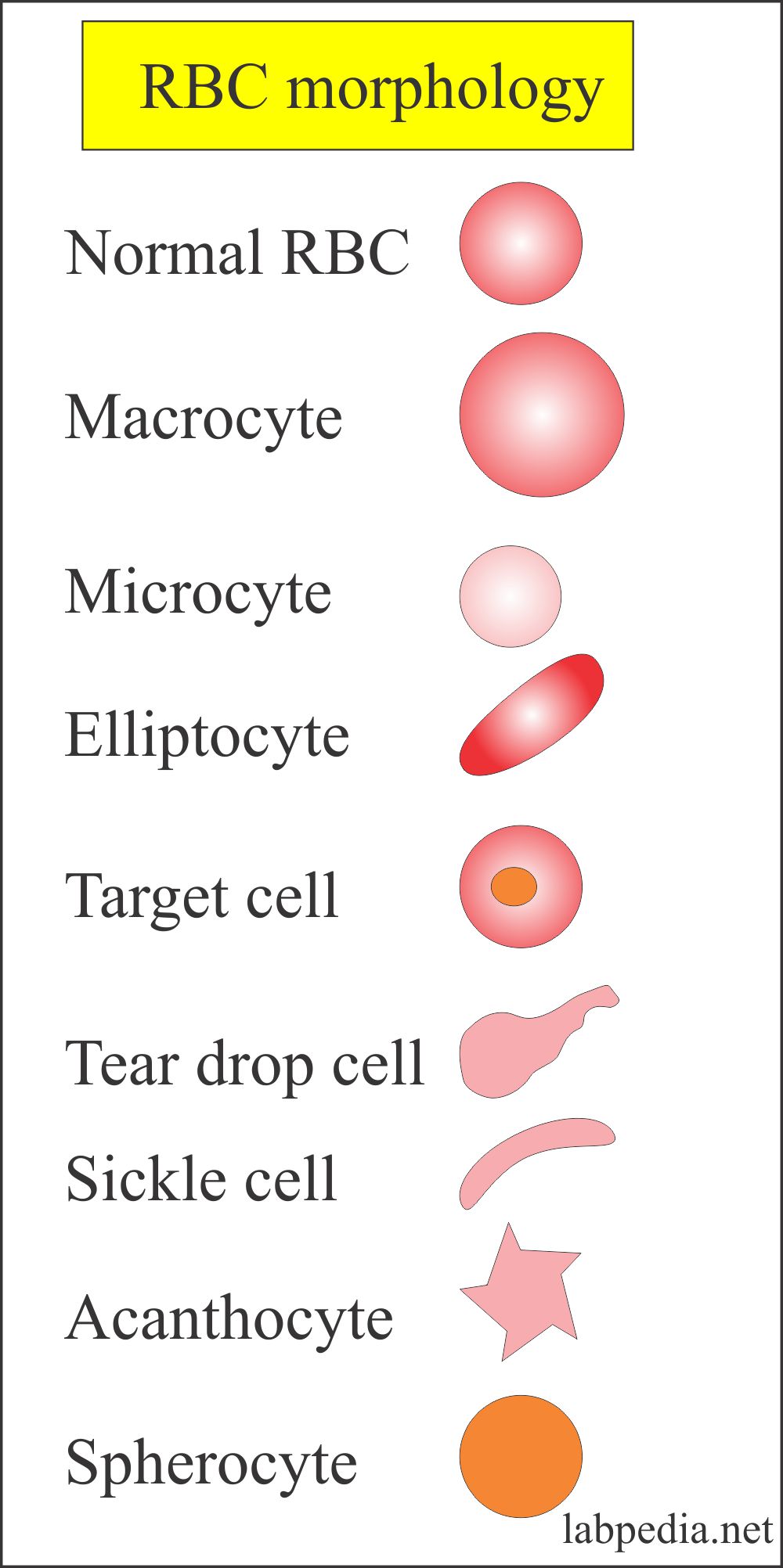
Blood Cell Morphology
https://www.labpedia.net/wp-content/uploads/2020/01/RBC-morphology-2.jpg

https://www.icsh.org › recommendations-for-standardization-of...
These International Council for Standarization of Haematology ICSH guidelines provide information on how to reliably and consistently report abnormal red blood cells white blood cells and platelets using manual microscopy Grading of abnormal cells nomenclature and a brief description of the cells are provided

https://onlinelibrary.wiley.com › doi › full
Currently there are two systems or methods of grading RBC M 2 4 11 While some laboratories report or grade the degree of morphologic abnormalities numerically plus from 1 to 4 others used descriptive terms such as slight

Rbc Morphology Grading Chart A Visual Reference Of Charts Chart Master

Rbc Morphology Grading Chart

Reporting And Grading Of Abnormal Red Blood Cell Morphology Artofit
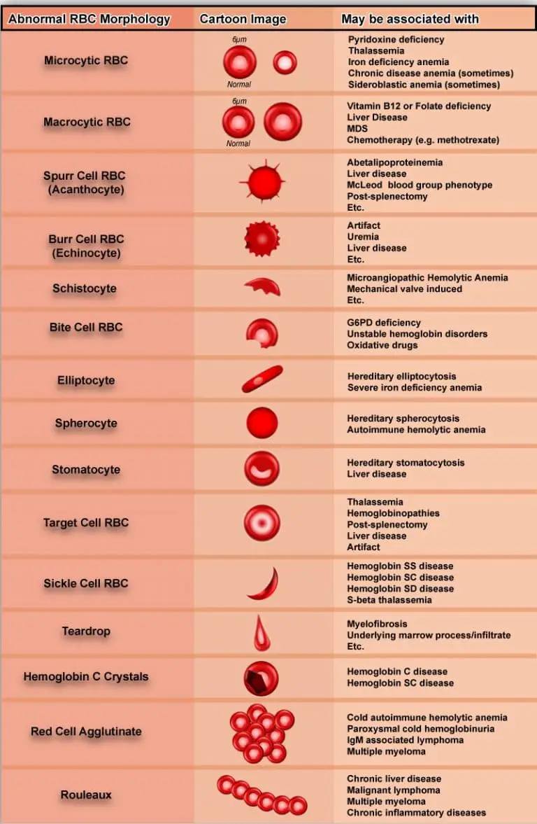
Variations In Red Blood Cell Morphology Size Shape Color And Inclusion Bodies
Red Blood Cell RBC Part 5 RBC Morphology Differential Diagnosis And Interpretations
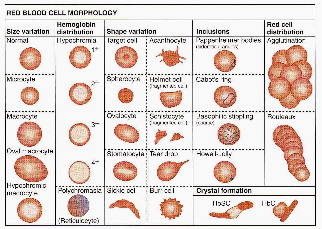
Medical Laboratory And Biomedical Science Red Blood Cell Morphology Abnormalities

Medical Laboratory And Biomedical Science Red Blood Cell Morphology Abnormalities
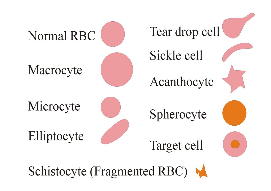
Red Blood Cell RBC Part 1 Morphology Peripheral Blood Smear Normal Picture Labpedia
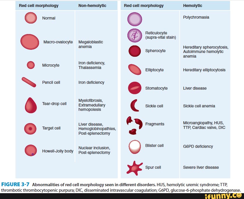
Sharing My Click Hole warning Blood Red Cell Morphology Non hemolytic Red Cell Morphology

Variations In Red Blood Cell Morphology Size Shape Color And Inclusion Bodies
Red Blood Cell Morphology Grading Chart - How are the WBC identified and classified granulocytes basophils monocytes lymphocytes WBC can also be identified and classified as to maturity i e mature cell or immature stage of development