Red Blood Cell Morphology Chart Hyperchromasia hyper over deep staining of the red cells with a lack of central pallor Polychromesia Poly many and chromesia color is present due to immature red blood cells which uptake Eosin Y Red Hb and Azure B Blue RNA They have a grayish blue color
Red blood cells erythrocytes are biconcave disks with a diameter of 7 8 microns which is similar to the size of the nucleus of a resting lymphocyte In normal red blood cells there is an area of central pallor that measures approximately 1 3 the diameter of the cell Download Table RED BLOOD CELL MORPHOLOGY GRADING CHART from publication Manual of HEMATOLOGY ResearchGate the professional network for scientists TABLE 1 uploaded by Gamal
Red Blood Cell Morphology Chart
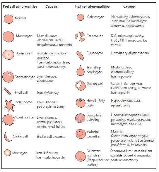
Red Blood Cell Morphology Chart
http://www.medical-labs.net/wp-content/uploads/2014/04/Red-cell-abnormalities.jpg

Red Blood Cell Morphology Grading
https://ai2-s2-public.s3.amazonaws.com/figures/2017-08-08/42bdc90e71c153741d0ff36fa13d0f318f3854ac/2-Figure1-1.png
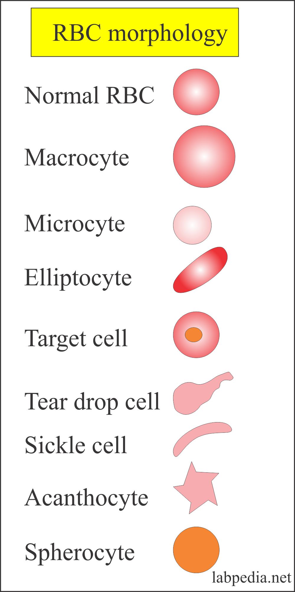
Blood Cell Morphology
https://www.labpedia.net/wp-content/uploads/2020/01/RBC-morphology-2.jpg
Explanation The red cell MCV is measured directly on cell counters this is true whatever method is employed to enumerate cells The counter is able to plot a red cell volume histogram and the mean is determined MCV can be calculated from the spun hematocrit as in option b Red blood cells constitute the primary cellular component in blood Red blood cells that are mature are biconcave discs which have no nucleus and are devoid of most cell organelles including the lysomes endoplasmic reticulum and mitochondria However a variety of abnormal erythrocyte morphology can be seen in a variety of medical conditions
Peripheral blood film from a healthy patient showing normal red blood cells with a central pallor about a third of the RBC diameter An overview of the common morphological changes in red blood cell and their clinicopathological significance The degree of size variation is indicated by the Red Cell Distribution Width RDW on the CBC The Mean Cell Volume MCV indicates the average size of red blood cells present in the specimen An increased MCV indicates that there are a significant number of macrocytes present
More picture related to Red Blood Cell Morphology Chart

Blood Cell Morphology
https://i.pinimg.com/originals/73/43/65/734365771b636fda23c4cec551c1368a.jpg
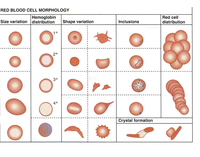
Red Blood Cell Morphology Printable Worksheet
https://cdn-useast.purposegames.com/images/game/bg/451/NcWMDqKhPrr.png?s=1400
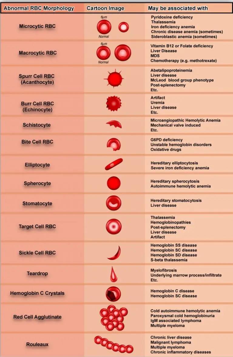
Variations In Red Blood Cell Morphology Size Shape Color And Inclusion Bodies
https://laboratoryinfo.com/wp-content/uploads/2015/04/Poikilocytosis-abnormal-rbc-shape-768x1177.jpg
How are the WBC identified and classified granulocytes basophils monocytes lymphocytes WBC can also be identified and classified as to maturity i e mature cell or immature stage of development Proper technique in the preparation and evaluation of a peripheral blood smear is essential in properly interpreting red blood cell morphology Ethylenediaminetetraacetic acid EDTA is the preferred anticoagulant for CBC testing and making peripheral blood smears
Red blood cell assessment is based on the following RBCs volume Normal MCV RBCs are called normocytic High MCV RBCs are called macrocytic Low MCV RBCs are called microcytic Hemoglobin contents Normal Hb and MCHC are called normochromic High MCHC RBCs are called hyperchromic Low MCHC RBCs are called hypochromic Red blood cell indices are Red blood cells have different morphological variations depending upon following type of inclusion bodies Howell Jolly bodies Small round cytoplasmic red cell inclusion with same staining characteristics as nuclei
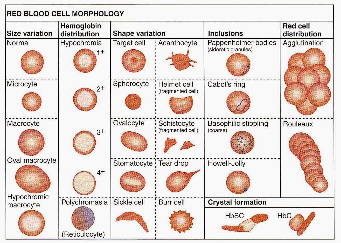
Medical Laboratory And Biomedical Science Red Blood Cell Morphology Abnormalities
http://3.bp.blogspot.com/--1Ifl-Yu46M/VR4z-qQmTVI/AAAAAAAAPsY/h0vzJP3KFPs/s1600/ii.jpeg

Red Blood Cell Morphology Chart 1488 The Best Porn Website
https://2.bp.blogspot.com/-0CgHJZd2w0A/WsSjVHP_6jI/AAAAAAAAEww/pzh59kz_d2M91XbJ3ES8DrPTJrpgP8BfgCLcBGAs/s1600/slide_8.jpg

https://faculty.ksu.edu.sa › sites › default › files
Hyperchromasia hyper over deep staining of the red cells with a lack of central pallor Polychromesia Poly many and chromesia color is present due to immature red blood cells which uptake Eosin Y Red Hb and Azure B Blue RNA They have a grayish blue color

https://askhematologist.com › blood-morphology
Red blood cells erythrocytes are biconcave disks with a diameter of 7 8 microns which is similar to the size of the nucleus of a resting lymphocyte In normal red blood cells there is an area of central pallor that measures approximately 1 3 the diameter of the cell

Vet Tech Infographic Canine Red Blood Cell Morphology Review Artofit

Medical Laboratory And Biomedical Science Red Blood Cell Morphology Abnormalities

Rbc Morphology Grading Chart
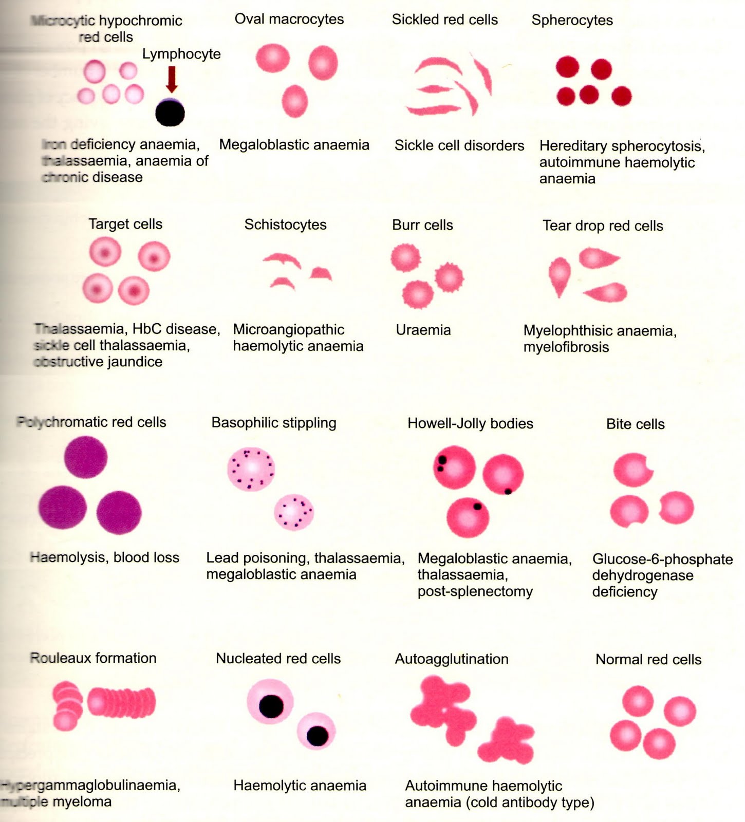
B L O O D ABNORMALITIES OF RED BLOOD CELLS

What Is Inside A Red Blood Cell Diagram
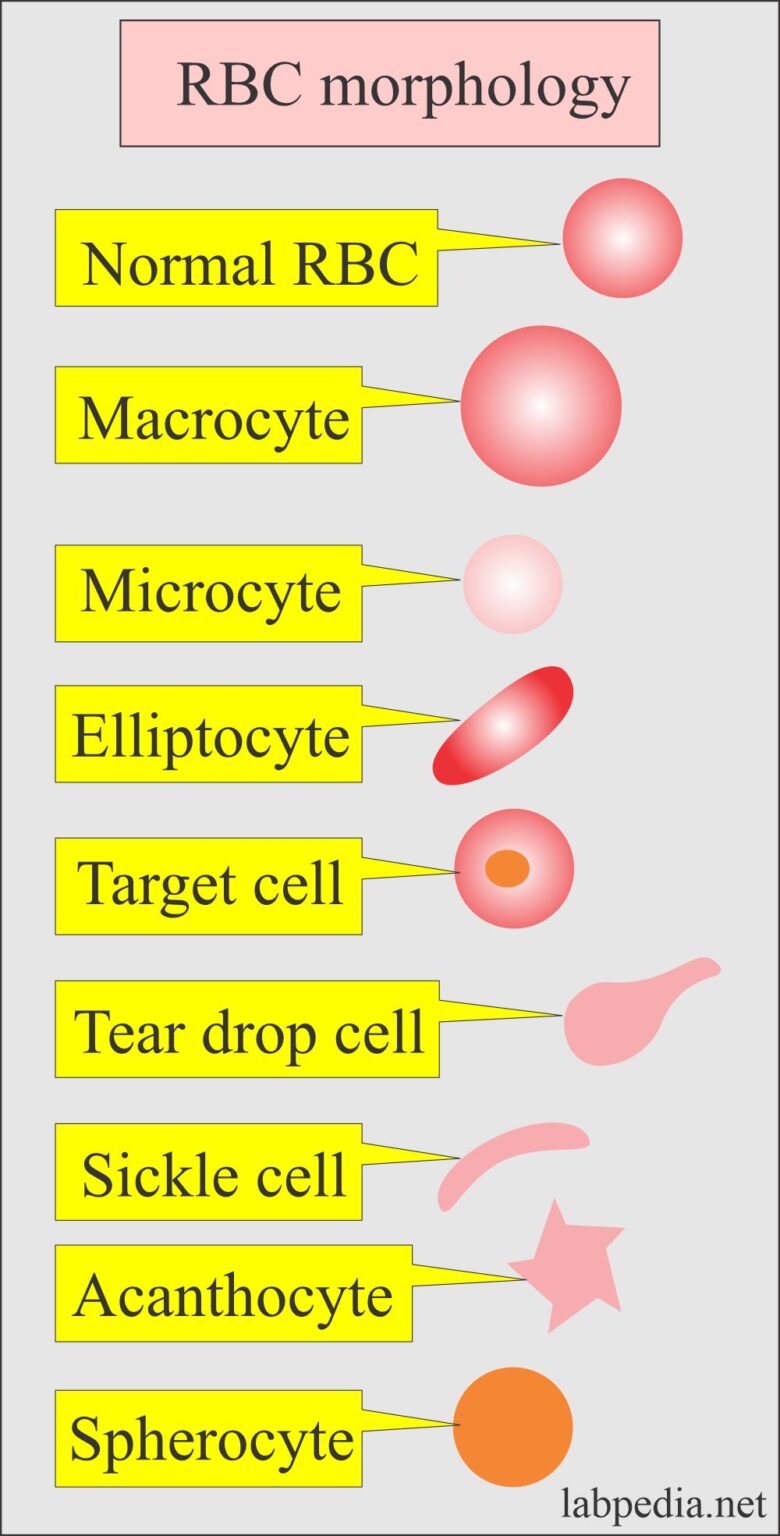
Complete Blood Count Red Blood Cell Morphology

Complete Blood Count Red Blood Cell Morphology
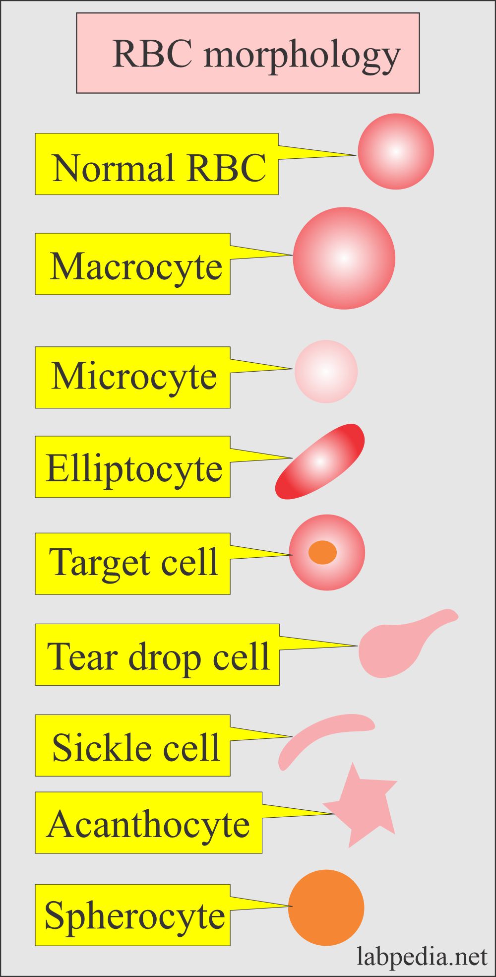
Complete Blood Count Red Blood Cell Morphology
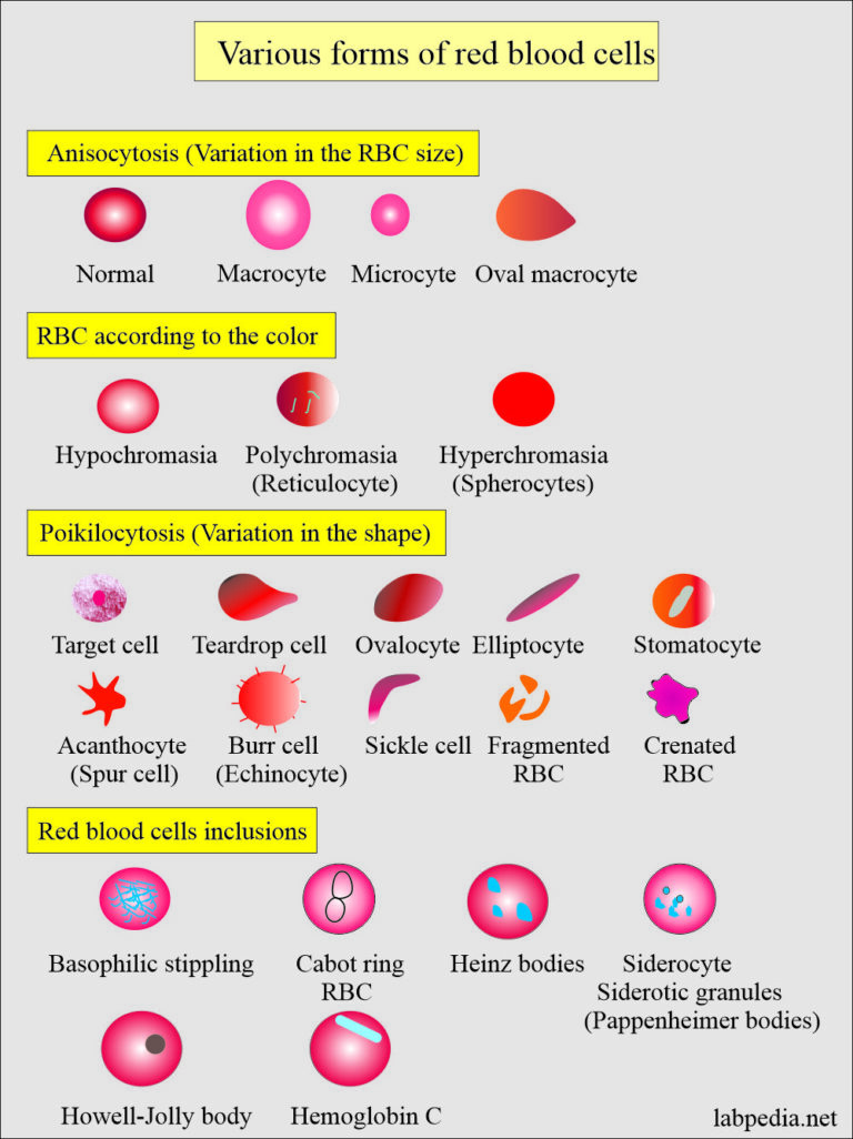
Complete Blood Count Red Blood Cell Morphology
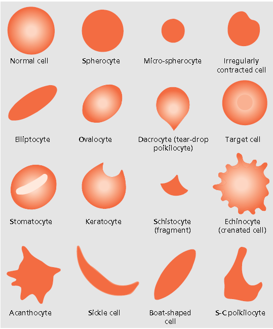
Medical Laboratory And Biomedical Science Blood Cell Morphology Guide
Red Blood Cell Morphology Chart - The degree of size variation is indicated by the Red Cell Distribution Width RDW on the CBC The Mean Cell Volume MCV indicates the average size of red blood cells present in the specimen An increased MCV indicates that there are a significant number of macrocytes present