Blood Vessel Elastic Tissue Smooth Muscle Tissue Chart Blood vessels namely arteries and veins are composed of endothelial cells smooth muscle cells and extracellular matrix including collagen and elastin These are arranged into three concentric layers or tunicae intima media and adventitia
Tunica intima TI endothelium subendothelial connective tissue and a wavy band of elastic fibers called the internal elastic lamina Tunica media TM up to 40 layers of smooth muscle cells an external elastic lamina is present in larger arteries Tunica adventitia TA thin layer of dense irregular connective tissue Get started with our blood vessel diagrams and artery and vein quizzes Stimulation of the alpha receptors produces contraction of the smooth muscles On the contrast stimulation of the beta receptors produces dilatation of the vessels Consequently this allows for sympathetic regulation of blood pressure
Blood Vessel Elastic Tissue Smooth Muscle Tissue Chart
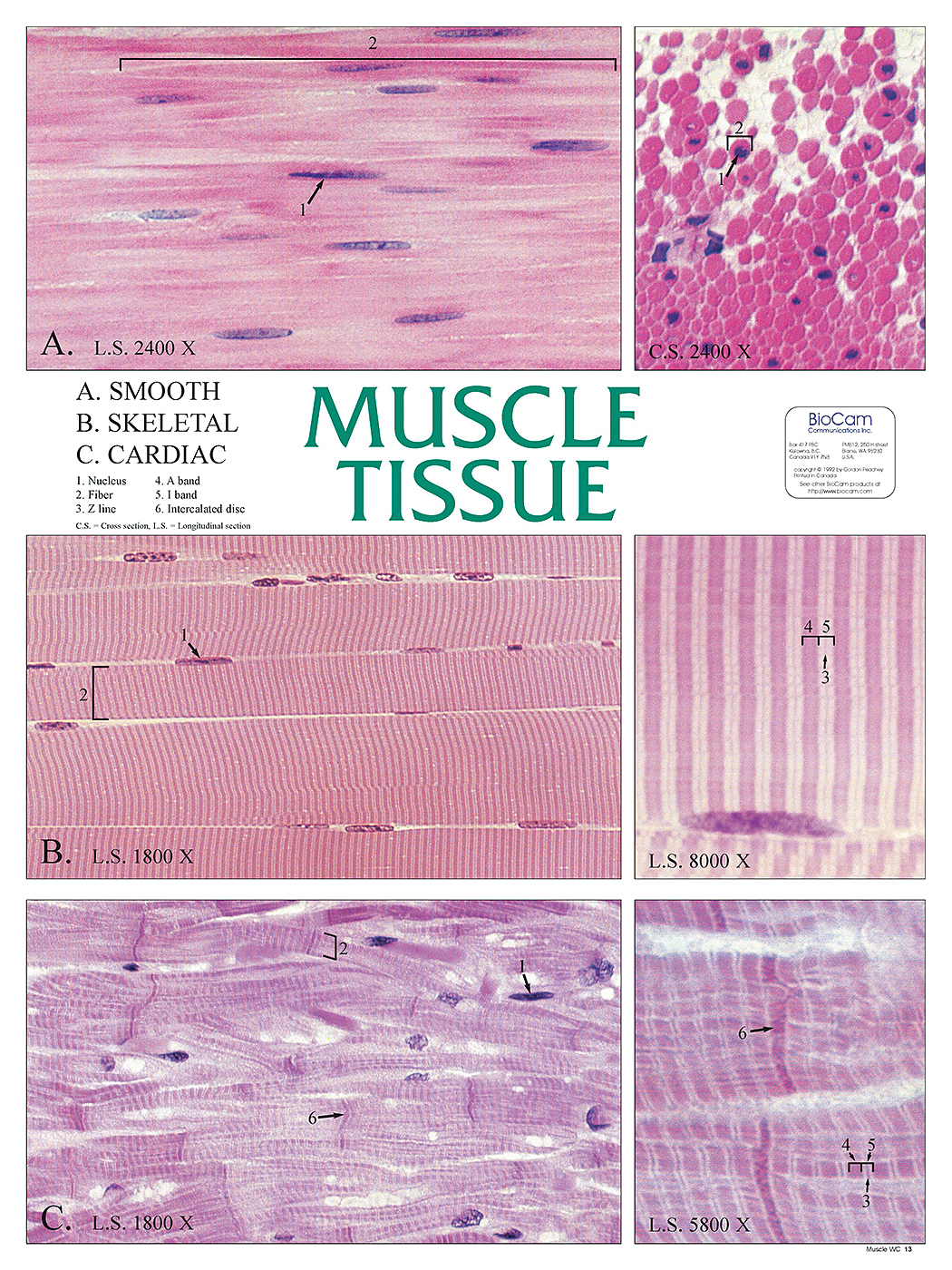
Blood Vessel Elastic Tissue Smooth Muscle Tissue Chart
https://www.flinnsci.ca/globalassets/flinn-scientific/all-product-images-rgb-jpegs/fb0370.jpg?v=a22d81e9a54a471097579a3b3dba23fa

Types Of Muscle Tissue Chart The Best Porn Website
https://i.pinimg.com/originals/99/7a/02/997a02954eac99ecc3152c808c86796f.jpg

Types Of Muscle Tissue Chart
https://i.pinimg.com/originals/da/4a/3c/da4a3c3803b9d8408796da86e80de01f.png
The middle and thickest layer of tissue of a blood vessel wall composed of elastic tissue and smooth muscle cells that allow the vessel to expand or contract in response to changes in blood pressure and tissue demand Start studying Chapter 20 Blood vessel anatomy Learn vocabulary terms and more with flashcards games and other study tools The arteriolar wall is composed of thin tunica intima with endothelial layers and subendothelial connective tissue tunica media with thin internal elastic lamina answer C and smooth muscle and tunica adventitia External elastic lamina is present only in muscular type arteries and the fenestrated elastic sheets are present in the
Types of Blood Vessels Arteries Elastic Muscular Arterioles Capillaries site of exchange with tissues Veins thinner walls than arteries contain less elastic tissue and fewer smooth muscle cells Venules Small veins Medium or large veins Blood Vessel Structure In general a blood vessel has three layer 1 Tunica interna or Compare and contrast the three tunics that make up the walls of most blood vessels Distinguish between elastic arteries muscular arteries and arterioles on the basis of structure location and function Describe the basic structure of a capillary bed from the supplying metarteriole to the venule into which it drains
More picture related to Blood Vessel Elastic Tissue Smooth Muscle Tissue Chart

Connective Tissue Structure And Anatomy Extracellular Matrix Elastic And Collagen Fibers
https://www.alamy.com/aggregator-api/download?url=https://c8.alamy.com/comp/2FM2W4G/connective-tissue-structure-and-anatomy-extracellular-matrix-elastic-and-collagen-fibers-blood-vessel-and-cells-lymphocyte-fibroblast-2FM2W4G.jpg

Tissue Raman Spectra Measured From Skeletal Muscle Tissue SM Fibrous Download Scientific
https://www.researchgate.net/publication/365152063/figure/fig3/AS:11431281107372329@1671031124343/Tissue-Raman-spectra-measured-from-skeletal-muscle-tissue-SM-fibrous-connective-tissue_Q640.jpg

Connective Tissue Classification Chart Porn Pics Sex Photos XXX Images Valhermeil
https://assets.fishersci.com/TFS-Assets/CCG/product-images/FS101349~p.eps-650.jpg
The middle and thickest layer of tissue of a blood vessel wall composed of elastic tissue and smooth muscle cells that allow the vessel to expand or contract in response to changes in blood pressure and tissue demand Resistance vessels Capacitance vessels store blood Thin walls The tunica media consists of layers of smooth muscle supported by connective tissue that is primarily made up of elastic fibers most of which are arranged in circular sheets Toward the outer portion of the tunic there are also layers of longitudinal muscle Contraction and relaxation of the circular muscles decrease and increase the diameter
The tunica media is the thickest layer in arteries and consists of smooth muscle layers and connective tissue that is made up of elastic fibers which appear wavy in histological slides Contraction and relaxation of the circular muscles decrease and increase the diameter of the vessel lumen respectively The arterioles can also constrict or dilate which varies their resistance to help distribute blood flow to the tissues Q A blood vessel with a few smooth muscle fibers and connective tissue and only a very thin tunica externa conducts blood toward the heart What type of vessel is this Answer A This is a venule Q An obese patient
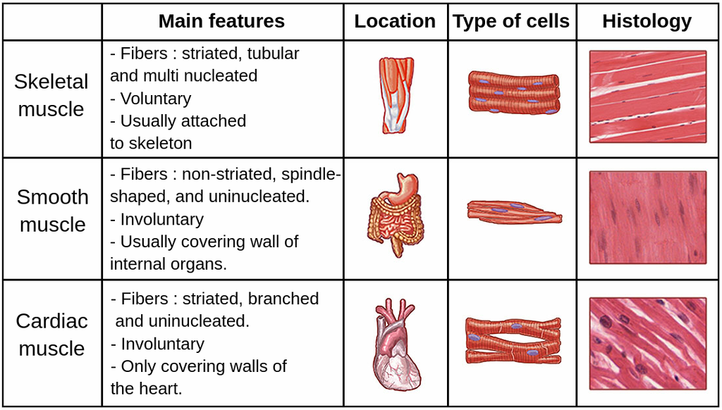
Skeletal Muscle Tissue Diagram Labeled Instituto The Best Porn Website
https://assets-global.website-files.com/6320bb946c79f421bd3b702f/63481613be75b52e69fcab8c_muscle_tissues.png
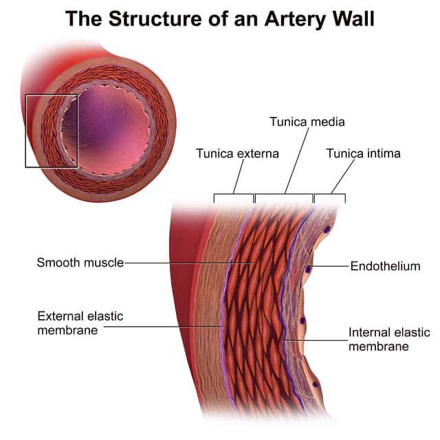
Blood Vessels TeachMePhysiology
https://teachmephysiology.com/wp-content/uploads/2021/04/Structure-of-an-Artery-Wall.jpg

https://radiopaedia.org › articles › histology-of-blood-vessels
Blood vessels namely arteries and veins are composed of endothelial cells smooth muscle cells and extracellular matrix including collagen and elastin These are arranged into three concentric layers or tunicae intima media and adventitia

https://histologyguide.com › figureview
Tunica intima TI endothelium subendothelial connective tissue and a wavy band of elastic fibers called the internal elastic lamina Tunica media TM up to 40 layers of smooth muscle cells an external elastic lamina is present in larger arteries Tunica adventitia TA thin layer of dense irregular connective tissue
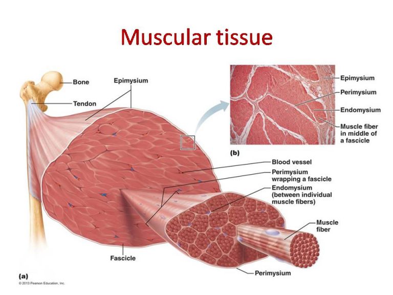
Proper Connective Tissue Areolar Adipose Reticular White Fibrous And Yellow Elastic Tissue

Skeletal Muscle Tissue Diagram Labeled Instituto The Best Porn Website

1 Structure Of Blood Vessel The Largest Blood Vessels Are Arteries Download Scientific

How To Identify Muscle Tissue
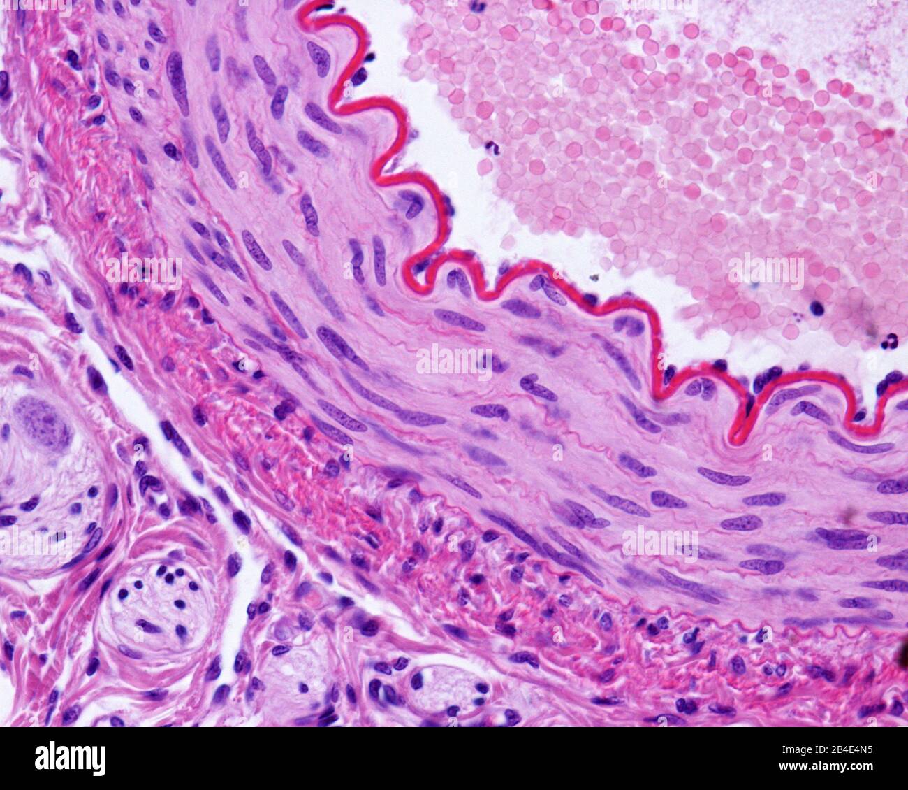
Light Micrograph Of A Cross sectioned Muscular Artery Showing A Thick And Wavy Internal Elastic
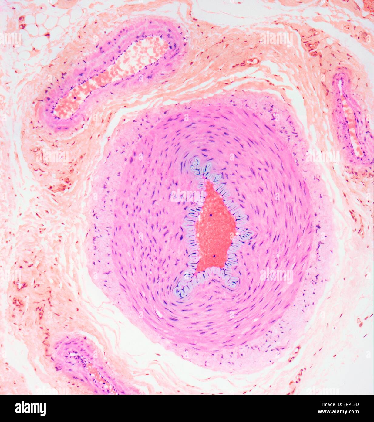
Blood Vessels Light Micrograph Of A Section Through Tissue Showing An Artery middle And A

Blood Vessels Light Micrograph Of A Section Through Tissue Showing An Artery middle And A
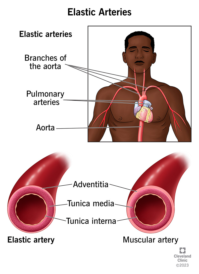
What Are Elastic Arteries
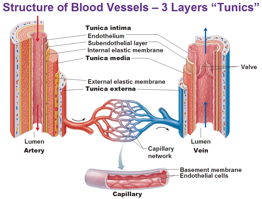
Blood Vessels
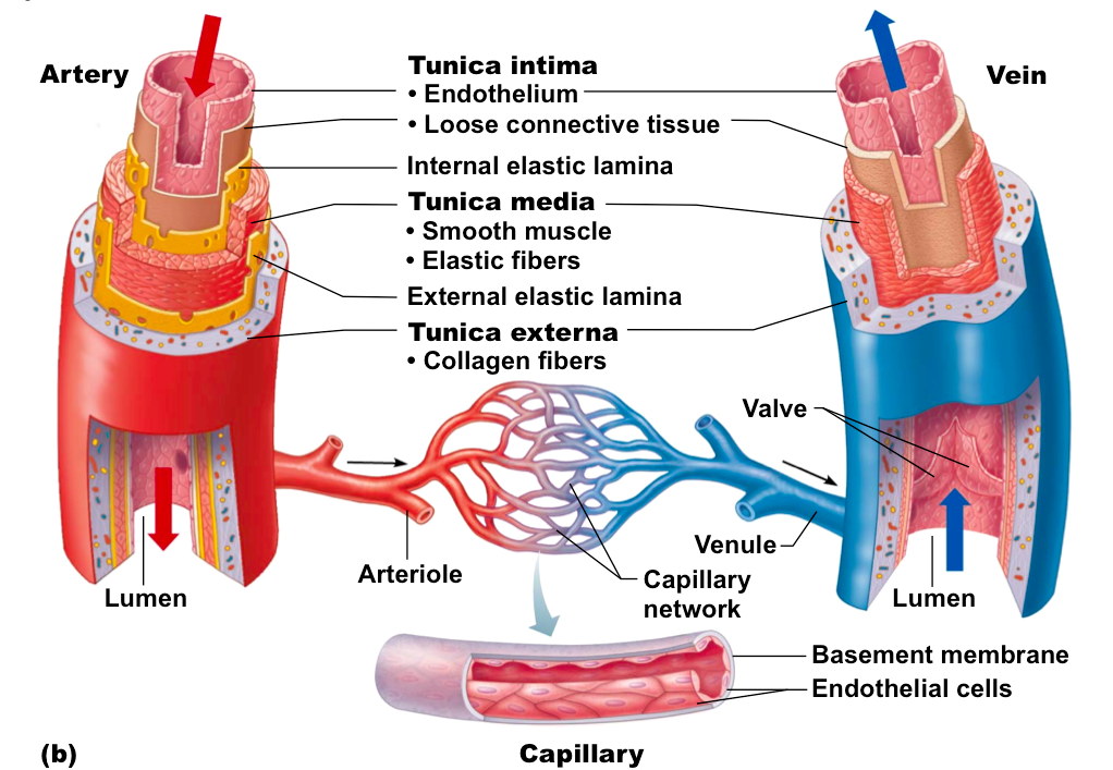
Blood Vessels Ms Gallagher s Classroom
Blood Vessel Elastic Tissue Smooth Muscle Tissue Chart - Types of Blood Vessels Arteries Elastic Muscular Arterioles Capillaries site of exchange with tissues Veins thinner walls than arteries contain less elastic tissue and fewer smooth muscle cells Venules Small veins Medium or large veins Blood Vessel Structure In general a blood vessel has three layer 1 Tunica interna or