Blood Pressure Chart Vena Cava Normal circulation with representative right and left cardiac pressures in mm Hg Representative right heart oxygen saturation 75 representative left heart oxygen saturation 95 Atrial pressures are mean pressures
Central venous pressure CVP describes the pressure of blood in the thoracic vena cava near the right atrium of the heart CVP reflects the amount of blood returning to the heart and the ability of the heart to pump the blood into the arterial system Central venous pressure is often used as an assessment of hemodynamic status particularly in the intensive care unit The central venous pressure can be measured using a central venous catheter advanced via the internal jugular vein and placed in the superior vena cava near the right atrium
Blood Pressure Chart Vena Cava

Blood Pressure Chart Vena Cava
https://www.ahajournals.org/cms/asset/6daedca8-f598-4767-b5aa-fceec6dc2f92/circimaging.122.014308.fig01.jpg
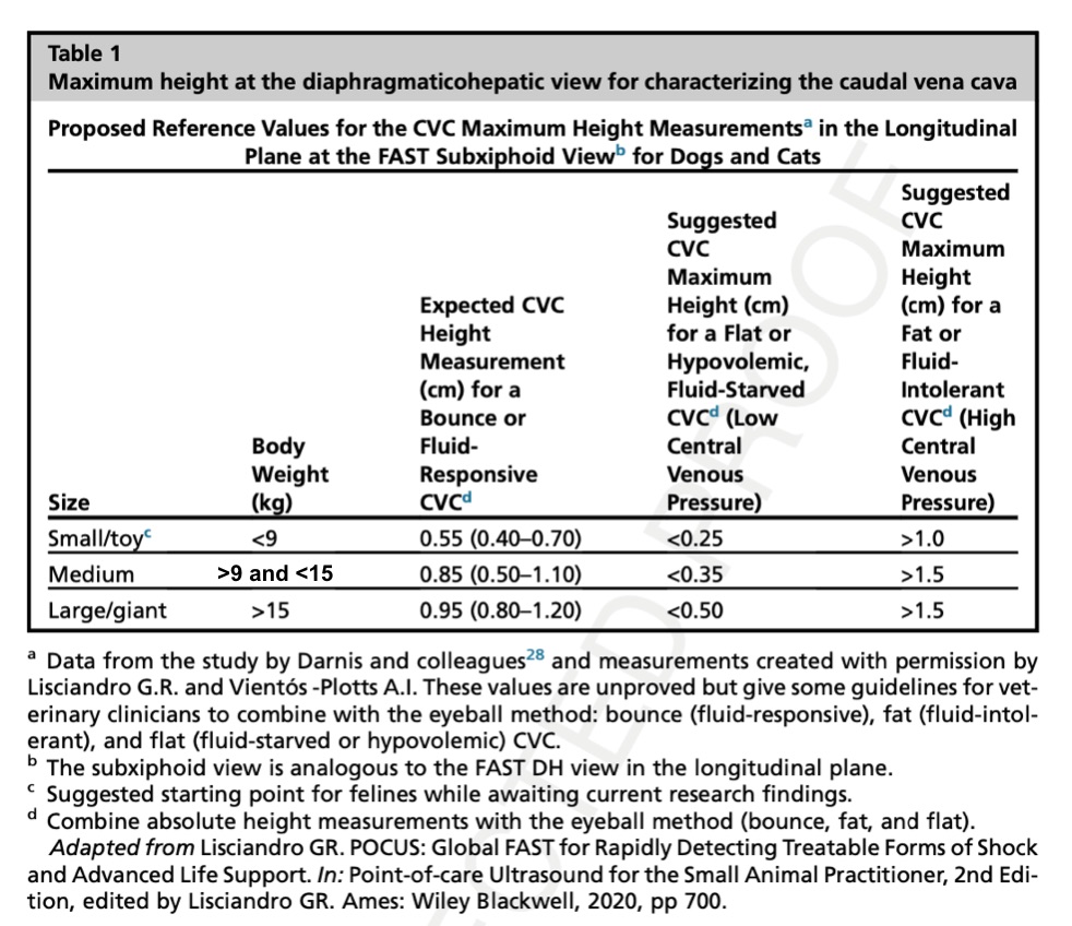
FASTVet Free Resources Caudal Vena Cava Maximum Height At The Subxiphoid DH View Measurement
https://fastvet.com/wp-content/uploads/2020/08/Caudal-Vena-Cava-Chart-Maximum-Height-8-28-2020.jpg
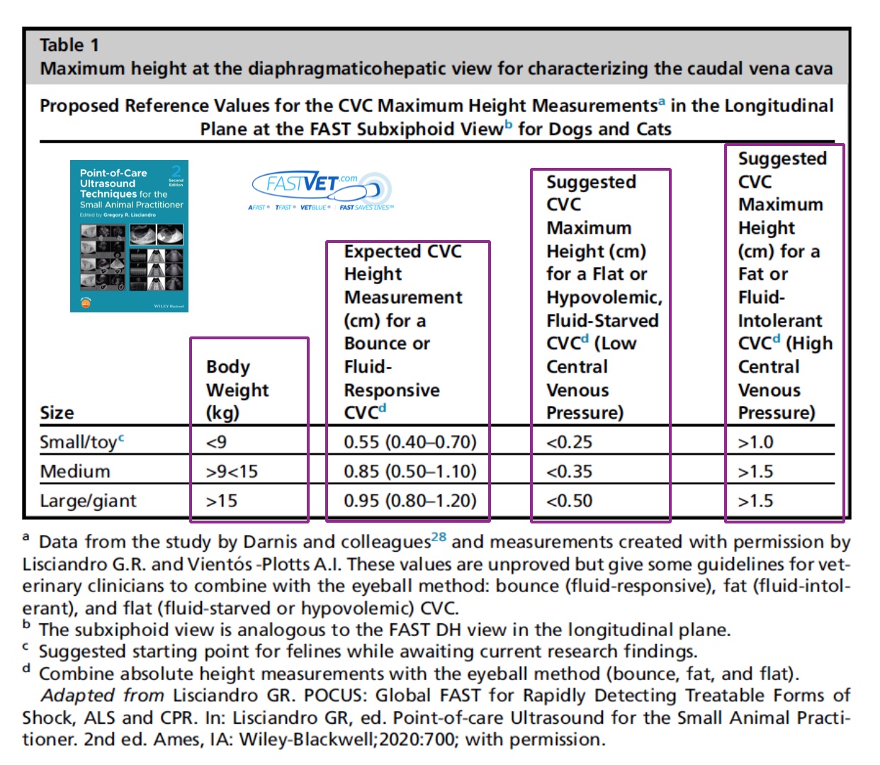
FASTVet Free Resources Caudal Vena Cava Maximum Height At The Subxiphoid DH View Measurement
https://fastvet.com/wp-content/uploads/2021/08/Updated-CVC-Chart-8-17-2021-IM-Blog-GBWE-JPEG.jpg
At a collapse of 60 70 the pressure is usually in the IVC 2 4mmHg With a collapse 10mmHg When the IVC is dilated and does not collapse IVC the pressures can reach 20 25mmHg IVC collaps registered with m mode Inferior vena caval IVC dimensions can be measured from the subcostal view on echocardiography If the inferior vena caval size is small less than 1 5 cm and collapses with inspiration the estimated right atrial pressure is below 5 mm Hg
Central venous pressure CVP is the most frequently used measure to guide fluid resuscitation in critically ill patients It is most often done via a central venous catheter CVC positioned in the right atrium or superior or inferior vena After the blood has passed through the capillary beds it enters the venules veins and finally the two main venae cavae singular vena cava that take blood back to the heart The flow rate increases again but is still much slower than the initial rate in the aorta
More picture related to Blood Pressure Chart Vena Cava

INFERIOR VENA CAVA New
https://medicoapps.org/wp-content/uploads/2018/10/1521376130.jpg
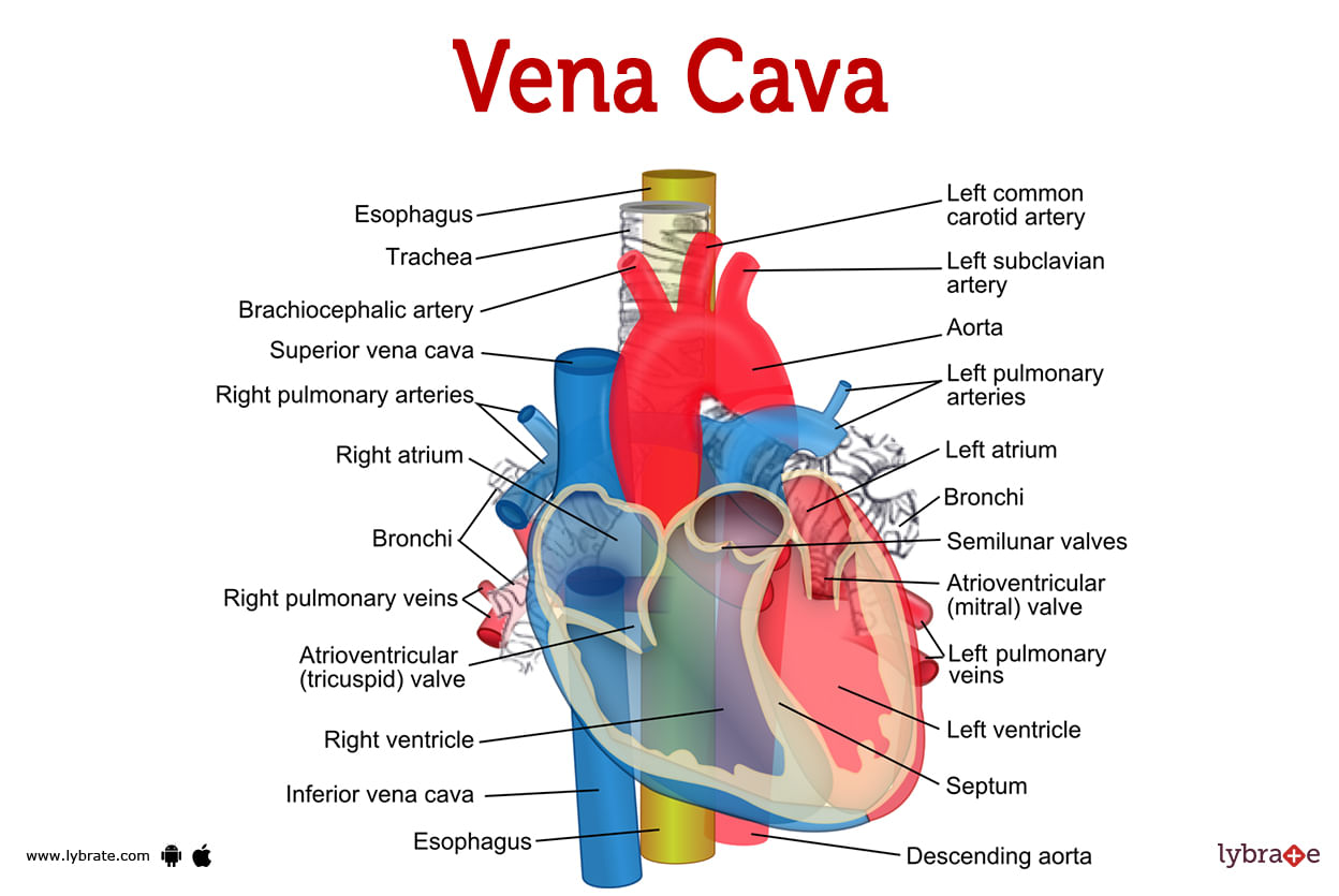
Vena Cava Human Anatomy Image Functions Diseases And Treatments
https://assets.lybrate.com/f_auto,c_limit,w_3840,q_auto/imgs/tic/enadp/image-of-the-vena-cava.jpg
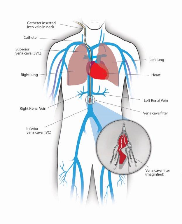
Vena Cava Filter Placement
https://vein.stonybrookmedicine.edu/sites/default/files/images/Patient Guide5retrieveFINAL2.jpg
Venous pressure values are commonly 5 mmHg in the right atrium and 8 mmHg in the left atrium Several measurements of venous blood pressure exist in various locations within the heart including central venous pressure jugular venous pressure and portal venous pressure The central venous pressure can be measured using a central venous catheter advanced via the internal jugular vein and placed in the superior vena cava near the right atrium A normal central venous pressure reading is between 8 to 12 mmHg This value is altered by volume status and or venous compliance
Vena cava Carries deoxygenated blood from the body back to the heart Pulmonary artery Carries deoxygenated blood from the heart to the lungs Pulmonary vein Carries oxygenated blood from Blood flows from the vena cava and pulmonary veins into the right and left atria respectively before flowing directly into the ventricles The ventricles fill with blood at a steadily decreasing rate until the ventricles pressure is equal to that in the veins

Vena Cava Occlusion Sequence From Base To Lowest Systolic Blood Download Scientific Diagram
https://www.researchgate.net/profile/Benjamin_Storm2/publication/371731082/figure/fig1/AS:11431281169361464@1687325424300/Vena-cava-occlusion-sequence-from-base-to-lowest-systolic-blood-pressure-The-top-line_Q640.jpg
Blood Pressure Chart Vine Spiral Bound
https://seventhsungallery.com/wp-content/uploads/2022/12/Vine-SSG.jpg

https://www.msdmanuals.com › professional › multimedia › table › norm…
Normal circulation with representative right and left cardiac pressures in mm Hg Representative right heart oxygen saturation 75 representative left heart oxygen saturation 95 Atrial pressures are mean pressures
https://nurseslabs.com › measuring-central-venous
Central venous pressure CVP describes the pressure of blood in the thoracic vena cava near the right atrium of the heart CVP reflects the amount of blood returning to the heart and the ability of the heart to pump the blood into the arterial system
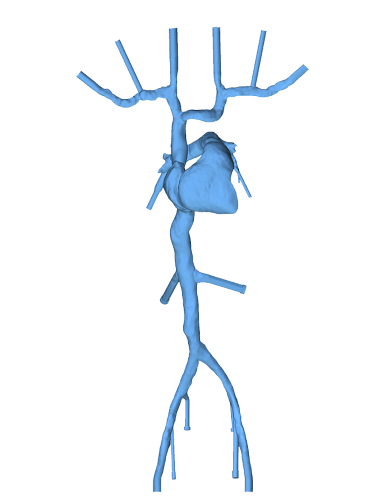
Vena Cava Heart Model Patient Specific United Biologics

Vena Cava Occlusion Sequence From Base To Lowest Systolic Blood Download Scientific Diagram
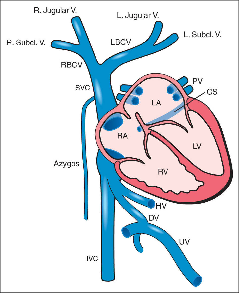
Superior Vena Cava Branches
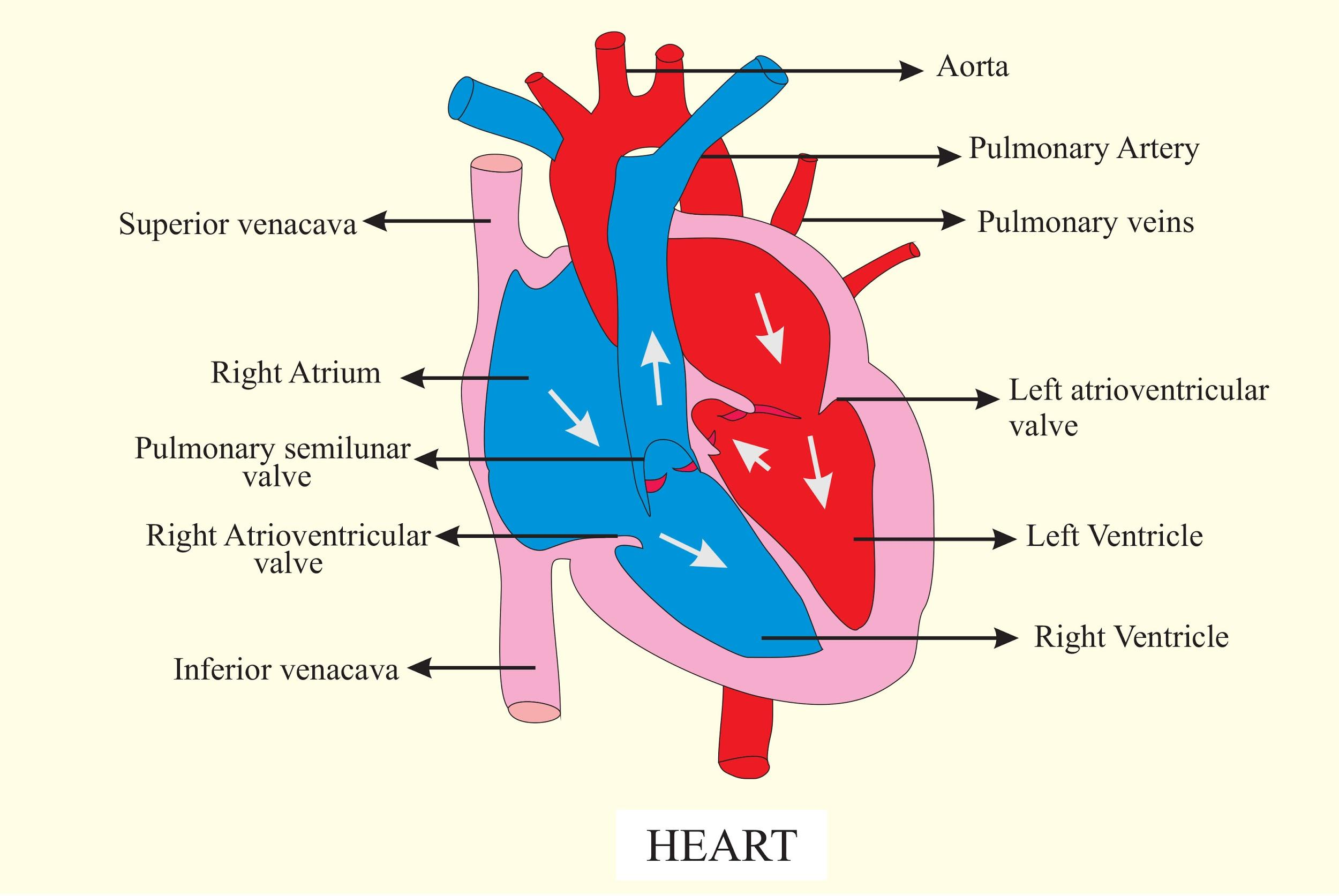
Cespuglio Frusta Fare Un Bagno Superior Vena Cava Function Bianco Come La Neve Evitare In Anticipo

Reference Chart For The Superior Vena Cava SVC Fraction Of Fetal Download Scientific

Solved 17 Superior Or Inferior Vena Cava Valve Valve Tru

Solved 17 Superior Or Inferior Vena Cava Valve Valve Tru

320 Vena Cava Vein Illustrations Stock Illustrations Royalty Free Vector Graphics Clip Art

Diagram Of Inferior Vena Cava Ultrasound 12321808 Vector Art At Vecteezy

Stage Classification Vena Cava Thrombus According To Staehler Own Download Scientific Diagram
Blood Pressure Chart Vena Cava - Objective To determine the most accurate predictor of central venous pressure among three point of care venous ultrasound techniques Design Cross sectional study Setting Medical ICU in an academic medical center