Blood Cells Histology Chart Rouleux Rouleaux is a normal finding in the thick part of a blood film but if present in the normal viewing area of the blood film it should be regarded as significant although not necessarily pathological Where marked rouleaux is present a cause should be sought consider physiological causes such as pregnancy reactive causes such as
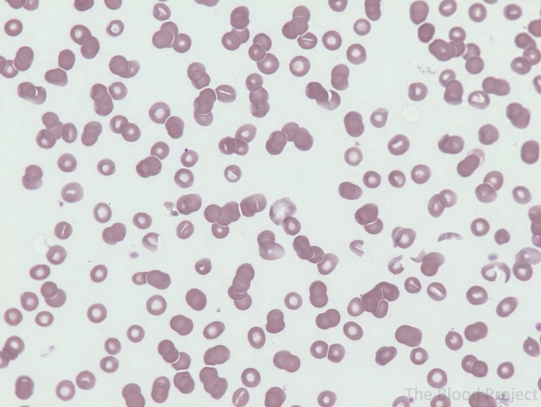
Rouleaux singular is rouleau are stacks or aggregations of red blood cells RBCs that form because of the unique discoid shape of the cells in vertebrates The flat surface of the discoid RBCs gives them a large surface area to make contact with Rouleaux formation in a 49 year old man with rheumatoid arthritis Note the stacked coin appearance of red cells in several parts of the field Overlap between red cells within the stacks may result in obscuring of central pallor 50x
Blood Cells Histology Chart Rouleux
Blood Cells Histology Chart Rouleux
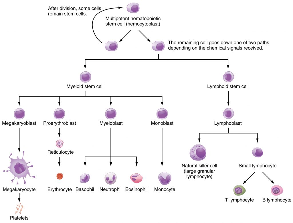
https://uta.pressbooks.pub/app/uploads/sites/36/2019/03/1902_Hemopoiesis-1024x778.jpg

Blood Cells Histology Labeled
https://o.quizlet.com/NTvFthUdHkDTgfFAs93UFg_b.png
![]()
Blood Cells Color Icons Set White Blood Cells Erythrocytes Platelets In The Blood Vessels
https://c8.alamy.com/comp/2WE2HTB/blood-cells-color-icons-set-white-blood-cells-erythrocytes-platelets-in-the-blood-vessels-vector-isolated-illustration-2WE2HTB.jpg
Cell Description Red blood cells are arranged into rows or linear chains appearing on top of one another in a coin stacking fashion The outlines of the the individual cells are usually seen 1 2 Cell Formation Can form naturally after blood is collected and allowed to sit for a Blood Blood Cells and Cellular Components Red Blood Cells Abnormal Rouleaux
The majority of shown erythrocytes form rouleaux due to the presence of an excessive amount of gamma globulins in the blood In this case gamma globulins are present in the form of pathological paraprotein the sample was taken from a patient with multiple myeloma Leucocytes are present in an adequate amount Rouleaux formation refers to the stacking of 4 or more red blood cells Red cell membranes have a negative charge zeta potential that causes red cells to repel each other
More picture related to Blood Cells Histology Chart Rouleux

Rouleaux Formation Of Red Blood Cells Stock Photo Image Of Smear Analysis 178095396
https://thumbs.dreamstime.com/b/rouleaux-formation-red-blood-cells-hematology-laboratory-rouleaux-formation-red-blood-cells-178095396.jpg
![]()
White Blood Cells Isometric Icon Vector Illustration Stock Vector Image Art Alamy
https://c8.alamy.com/comp/2WKB3TA/white-blood-cells-isometric-icon-vector-illustration-2WKB3TA.jpg

Rouleaux formation Medicine Science And More
http://medicine-science-and-more.com/wp-content/uploads/rouleaux-formation.jpg
A This image demonstrates rouleaux formation RBC forming stacks of variable length in a cat with a monoclonal gammopathy due to multiple myeloma The blue background is also a reflection of the high protein in the sample 911 5 g dL This occurs when red blood cells stack on top of each other when seen on a peripheral blood film It is the blood film equivalent of a raised ESR An article from the haematology section of GPnotebook Rouleaux formation
The stacking of cells rouleaux formation facilitates the rate of red cell sedimentation a phenomenon that may be seen on a peripheral smear The appearance of rouleaux may be artificially caused by a poor preparation of the smear or by viewing the slide in a thickened area The rouleaux formation is a phenomenon of turbulent blood flow in an area of blood stasis Although the rouleaux formation is a common ultrasonography phenomenon and sometimes is not considered a clinical effect it is important to note that it is usually found at a lesion with a proximal venous obstruction such as venous stenosis or deep
Rouleaux The Blood Project
https://www.thebloodproject.com/wp-content/uploads/2021/09/Rouleaux1-776x584.png
Blood Cells Seamless Pattern Hand Drawn Erythrocytes Leukocytes And Platelet Scientific
https://c8.alamy.com/comp/2P231XD/blood-cells-seamless-pattern-hand-drawn-erythrocytes-leukocytes-and-platelet-scientific-biology-illustration-in-sketch-style-2P231XD.jpg
https://www.haematologyetc.co.uk › index.php
Rouleaux is a normal finding in the thick part of a blood film but if present in the normal viewing area of the blood film it should be regarded as significant although not necessarily pathological Where marked rouleaux is present a cause should be sought consider physiological causes such as pregnancy reactive causes such as

https://en.wikipedia.org › wiki › Rouleaux
Rouleaux singular is rouleau are stacks or aggregations of red blood cells RBCs that form because of the unique discoid shape of the cells in vertebrates The flat surface of the discoid RBCs gives them a large surface area to make contact with

Red Blood Cells And White Blood Cells Diagram

Rouleaux The Blood Project
Blood Cells Seamless Pattern Hand Drawn Erythrocytes Leukocytes And Platelet Scientific
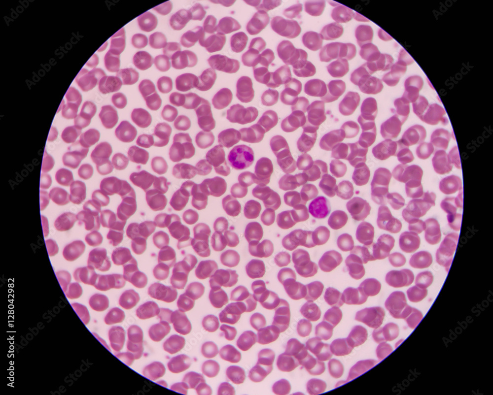
Blood Smear Shows Rouleaux Formation And White Blood Cell Stock Photo Adobe Stock
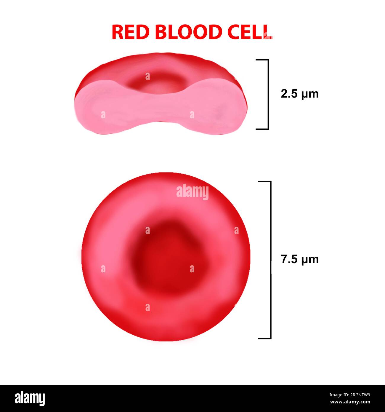
Red Blood Cells Structure Diagram
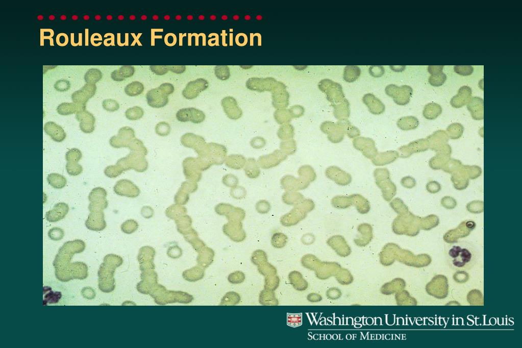
PPT Peripheral Blood Smears PowerPoint Presentation Free Download ID 3969425

PPT Peripheral Blood Smears PowerPoint Presentation Free Download ID 3969425

Peripheral Blood Smear Results Revealed A Red Blood Cell Rouleaux Download Scientific Diagram
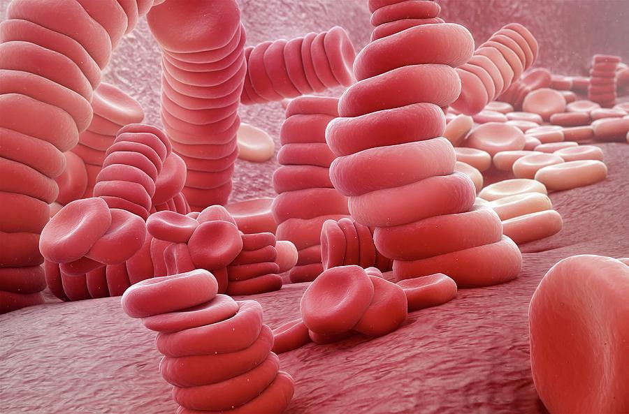
Red Blood Cell Rouleaux Photograph By Scientificanimations Science Photo Library Pixels

Figure 1 From The Decrease Of Rouleaux Formation Of Red Blood Cells In Healthy Human By Water
Blood Cells Histology Chart Rouleux - When true rouleaux is present it can be observed in the optimal smear viewing area at a magnification of 1000X It is also readily observable at a magnification of 400X Switching to this lower magnification will help provide confirmation when rouleaux is suspected on a blood smear